No products
To be determined
Shipping
0,00 €
Total
Welcome to the Lambey website
Select your country
Blog Categories
Latest Posts


« a horse that may not yet limp but is moving unusually or possibly not performing as well at work must be examined… »
In order to be able to move freely, horses need healthy synovial joints, which:
A key component of the synovial joint is the cartilage, a well-structured connective tissue made of cells knowns as chondrocytes, and an extracellular matrix of collagen and proteoglycanes (containing glycosaminoglycans such as hyaluronic acid) which acts like a sponge. It flattens when weight is placed on the limb, and fills with synovial fluid during the swing phase of the gait. The integrity of the peripheral soft tissues (capsule and ligaments) offers essential stability for all components of the joint.
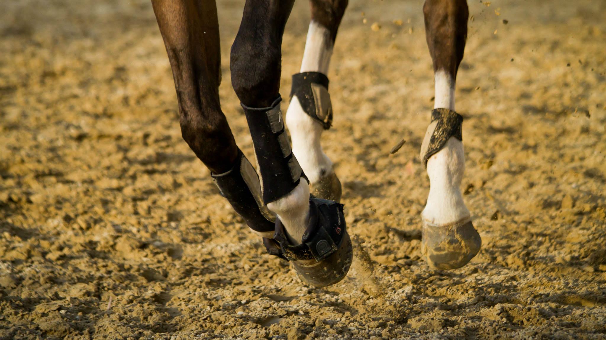 |
This complex anatomical structure is vulnerable to various problems with multiple causes. If not due to a deformation or illness such as osteochondrosis, it is often the handler who, forcing his mount to undertake repeated and sometimes unsuitable movements, places abnormal biomechanical stress on one of the components of the joint. The work of a sport horse is hugely demanding on its joints and, depending on the discipline, some are subject to more biomechanical stress than others and are therefore more vulnerable to degenerative joint disease (DJD). |
All types of joint trauma will trigger the release of various inflammatory mediators (e.g. metalloproteinases, prostaglandins, free radicals, interleukin-1) which can affect the integrity of the cartilage. Progressive cartilage damage in turn releases other inflammatory mediators, creating and sustaining a vicious degenerative cycle.
Clinically, the inflammation caused by DJD results in swollen joints, pain, limited range of movement and, in the rider's trained eye, a limp. Depending on which joint component is affected, DJD goes by different names, such as capsulitis, synovitis (usually with a sprain), arthritis or osteoarthritis when the cartilage and/or subchondral bone is involved. If the horse has a limbs deformation, osteochondrosis, or suffers a single major trauma to a joint, DJD can develop quite rapidly; on the other hand, in sport horses subjected to repeated mechanical stress the symptoms may only appear after several years of competing. The limbs are naturally the most affected, but the spine, especially the cervical spine, is also involved.
An early and accurate diagnosis is key to successful treatment. After a thorough physical examination, sometimes using an accelerometer-based system such as the “Lameness Locator™” to detect any limp, with the help of semiological anaesthesia to locate the site, the veterinarian may use different medical imaging techniques. However, none of these can provide a particularly good view of the cartilage, for which the go-to technique involves inserting a small camera called an arthroscope into the joint.
DJD is a progressive degenerative condition which cannot be reversed once the cartilage has worn away. Effective management therefore means preventing onset or stopping progression and managing the pain. Preventing the DJD in the first place is of course the ideal solution. This can be done by:
Although such products are safe in horses and pain relief has been achieved with a course of harpagophytum, their chondral protection claims need further study to assess their actual therapeutic effects. Chondral protection means the ability to prevent cartilage damage and stimulate repair, which is far from being proven for most products on the market. During the acute phase of most cases of DJD, rest combined with systemic non-steroidal anti-inflammatories is sufficient to minimise the symptoms and control the pain. Another effective way of putting a rapid stop to the degenerative vicious cycle is joint injections (corticosteroids, hyaluronic acid, interleukin 1 RAP, stem cells). Of these options, the most effective is corticosteroids, but care should be taken to observe the minimum washout time before any competition, which can vary depending on the molecule used. Involvement of the subchondral bone during excessive osteoclastic activity, diagnosed using x-rays, is an indication for bisphosphonate therapy (tiludronic or clodronic acid).
The surgical options vary depending on whether the DJD affects a joint with a small or large range of motion. For joints with a small range of motion (pastern, intertarsal joints), arthrodesis can be used to fuse the joint, preventing any movement and thus alleviating the pain. For joints with a large range of motion (fetlock, stifle, carpal), arthroscopy or arthrotomy is required to debride the damaged cartilage, remove any fragments, repair the fractures and possibly, in the future, resurface the joint and conserve the joint range of movement. There is debate over the usefulness of stem cell injections, and this type of treatment cannot ever regenerate the defect. The hope is that this can be achieved with an autologous cartilage graft (using cells taken from the same horse) onto the osteoarthritis lesions. Cartilage cells harvested from the patient are grown in a 2D/3D culture before being reimplanted into the lesion as small beads of cartilage (chondrobeads). This is one of the topics being researched by the Equine Sports Rehabilitation and Medicine Research Group at the Lyon Veterinary School, working in collaboration with the team under Dr V. Tieng at Geneva University Hospital. It is a good example of a “one health” approach involving the transfer of technology from human medicine to veterinary medicine and vice versa, with the ultimate goal of improving the well-being of both horse and rider.
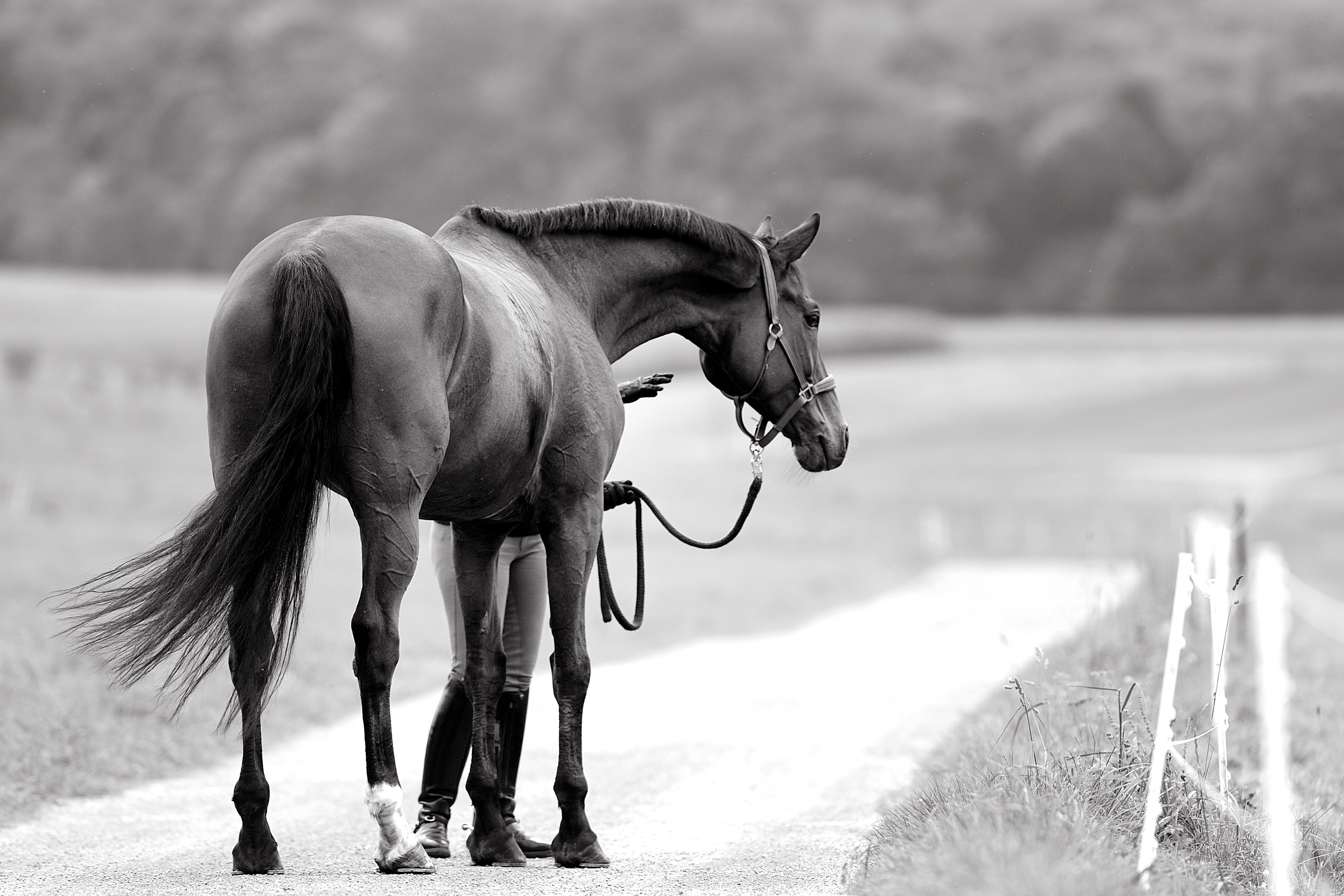 |
A horse could be exposed to abnormal joint stress at any point of its sporting career, resulting in DJD. The rider has a duty to recognise, and the veterinarian has a duty to diagnose, this condition as quickly as possible to halt the process before any damage to the cartilage can occur. DJD is reversible in the form of capsulitis, synovitis and even if there are joint fragments, but a destroyed cartilage will never regenerate and the repair surgery for this is still at the experimental stage. Every effort must therefore be taken to prevent such damage from occurring, and although veterinary research needs to focus on medical imaging and regenerative techniques, a horse that may not yet limp but is moving unusually or possibly not performing as well at work must be examined. When it comes to DJD, prevention is cure! |